44 microscope diagram with labels
Labeled Parts Compound Microscope [4THWBQ] A compound light microscope is a type of light microscope that uses a compound lens system meaning it operates through two sets of lenses to magnify the image of a specimen Two different compound light microscope models with their parts labeled Leica DM1000 Fluorescence Filter - Blue - 11513828 Compound Microscopes Defining Features Image 1 ... Scanning Electron Microscope (SEM) - Diagram, Working Principle ... Definition. Scanning electron microscope is a classification of electron microscope that uses raster scanning to produce the images of a specimen by scanning using a focused electron beam on the surface of the specimen. An SEM creates magnified images of the specimen by probing along a rectangular area of the specimen with a focused electron beam.
anatomysystem.comAnatomy System - Human Body Anatomy diagram and chart images ... Cell Picture With Labels Image Diagram - Cell Picture With Labels Image Chart - Human anatomy diagrams and charts explained. This anatomy system diagram depicts Cell Picture With Labels Image with parts and labels. Best diagram to help learn about health, human body and medicine.

Microscope diagram with labels
› techniques › multi-photonMultiphoton Microscopy | Nikon’s MicroscopyU This is a valuable enhancement to the capability of the conventional microscope since ultraviolet wavelengths below approximately 300 nanometers are very problematic for regular microscope optics. Higher-order non-linear effects, such as four-photon absorption, have been experimentally demonstrated, although it is unlikely that these phenomena ... Compound Microscope - Diagram (Parts labelled), Principle and Uses See: Labeled Diagram showing differences between compound and simple microscope parts Structural Components. The three structural components include. 1. Head. This is the upper part of the microscope that houses the optical parts. 2. Arm . This part connects the head with the base and provides stability to the microscope. Bright-field microscope (Compound light microscope) - Diagram (Parts ... A few applications of the bright-field microscope include: Used to observe, analyze, and study plant cells. Used to view, magnify, and study about animal cells. Used to clearly study the morphologies of bacterial, and viral organisms. Also used in the study of parasites like paramecium. It finds use in agricultural laboratories to study soil ...
Microscope diagram with labels. rsscience.com › stereo-microscopeParts of Stereo Microscope (Dissecting microscope) – labeled ... Labeled part diagram of a stereo microscope Major structural parts of a stereo microscope. There are three major structural parts of a stereo microscope. The viewing Head includes the upper part of the microscope, which houses the most critical optical components, including the eyepiece, objective lens, and light source of the microscope. Labeled Parts Microscope Compound [VX97ET] Home » Instrumentation » Compound microscope- definition, labeled diagram, parts, uses, Last Updated on February 24, 2020 by Sagar Aryal Image 3: A compound microscope with a corresponding label of the different parts Microscope Diagram Labeled, Unlabeled and Blank It comes with a wide body and base It is the standard microscope that is used ... › createJoin LiveJournal Password requirements: 6 to 30 characters long; ASCII characters only (characters found on a standard US keyboard); must contain at least 4 different symbols; Difference between Simple and Compound Microscope A simple microscope employs a single lens while a compound microscope employs more than one lens to achieve higher order of magnification. A simple microscope is used for basic magnification of specimens and is generally used in the fields of soil study, schools, dermatology among others. Compound microscope, on the other hand, are used in more ...
microscopewiki.com › simple-microscopeSimple Microscope - Diagram (Parts labelled), Principle ... Oct 01, 2022 · The arm of a simple microscope is made of metal and it connects the base of the microscope with the lens tube that houses the eyepiece. Stage . This is a rectangular metal plate with a hole in the middle and is attached to the body of the microscope. The hole in the middle is called the aperture and it allows the light to fall on the specimen. en.wikipedia.org › wiki › Wikipedia:Citation_neededWikipedia:Citation needed - Wikipedia If someone tagged your contributions with a "Citation needed" tag or tags, and you disagree, discuss the matter on the article's talk page.The most constructive thing to do in most cases is probably to supply the reference(s) requested, even if you feel the tags are "overdone" or unnecessary. Spleen histology: Location, functions, structure | Kenhub Spleen histology slide (labeled) The spleen is a fist sized organ located in the left upper quadrant of the abdomen.It is the largest lymphoid organ and thus the largest filter of blood in the human body.The spleen has a unique location, embryological development and histological structure that differs significantly from other lymphoid organs.. Special histological features define several ... Compound microscope - BiochemGems Figure: A labeled diagram of a compound microscope. Image formed by a compound microscope. The objective lens forms a true, inverted picture while the eyepiece functions as a basic magnifier that does not re-invert and generates a virtual image. The image always ends up inverted from the original. If we move the sample to the left, it will move ...
Compound Microscope Parts Labeled [REJA3G] This is an online quiz called Microscope Parts Diagram This is an online quiz called Microscope Parts Diagram. The main parts of a microscope that are easy to identify include: Head: The upper part of the microscope that houses the optical elements of the unit They explore various websites, label the parts of a microscope on a worksheet, … Structure of Cell: Definition, Cell Theory, Plant and Animal Cells - Embibe Cells in Latin means 'little rooms'. This name was given by Scientist Robert Hooke in the year \(1665\) who discovered the cell using a self-designed microscope. While studying a thin slice of cork (a substance that is obtained from the bark of a tree), Robert Hooke saw that the cork resembled the structure of a honeycomb consisting of many ... Parts Microscope Compound Labeled [TX8NJI] Labeled Diagram of a Compound Microscope The Microscope Parts and Use Name:_____ Period:_____ Historians credit the invention of the compound microscope to the Dutch spectacle maker, Zacharias Janssen, around the year 1590 T-Mount A standard adapter for mounting 35mm cameras to microscopes … Skin: Cells, layers and histological features | Kenhub The organ constitutes almost 8-20% of body mass and has a surface area of approximately 1.6 to 1.8 m2, in an adult. It is comprised of three major layers: epidermis, dermis and hypodermis, which contain certain sublayers. Owing to variations in height and weight, the surface area of the skin may vary based on these parameters.
Simple Microscope - Diagram (Parts labelled), Principle ...
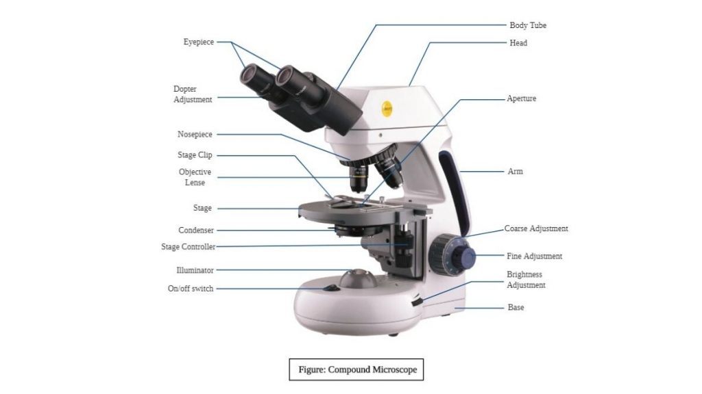
Binocular Microscope Anatomy - Parts and Functions with a Labeled Diagram The important non-optical parts of the light compound microscope are the body tube or head, arm or frame, fine adjustment, coarse adjustment, nose piece, stage, and base. Now, I will describe all these non-optical parts of the light compound microscope with the labeled diagrams. The body tube of the microscope

Compound Microscope- Definition, Labeled Diagram, Principle ...
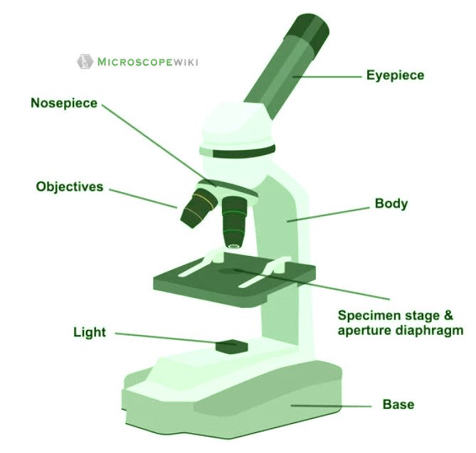
Microscope Parts, Types & Diagram | What is a Microscope? The essential parts include the head, base, arms, lenses, and lights. In diagrams, one would see the head always located at the top of the microscope while the base is at the bottom. The arms of a ...

Microscope Diagram Labeled, Unlabeled and Blank | Parts of a ...
quizlet.com › 231339270 › chapter-1-flash-cardsChapter 1 Flashcards | Quizlet Study with Quizlet and memorize flashcards containing terms like Physiology is to _____ as anatomy is to _____., Which of the following is arranged in correct order from the simplest to the most complex?, The study of tissues with a microscope is called _____. and more.
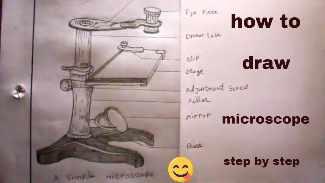
How TO Draw simple microscope step by step/simple microscope drawing/for science project
Electron Microscope Principle, Uses, Types and Images (Labeled Diagram ... Ans: A light microscope has a low resolving power (0.25µm to 0.3µm) while the electron microscope has a resolution power about 250 times higher than the light microscope at about 0.001µm. Similarly, a light microscope has a magnification of 500X to 1500x while the electron microscope has a much higher magnification of 100,000X to 300,000X.

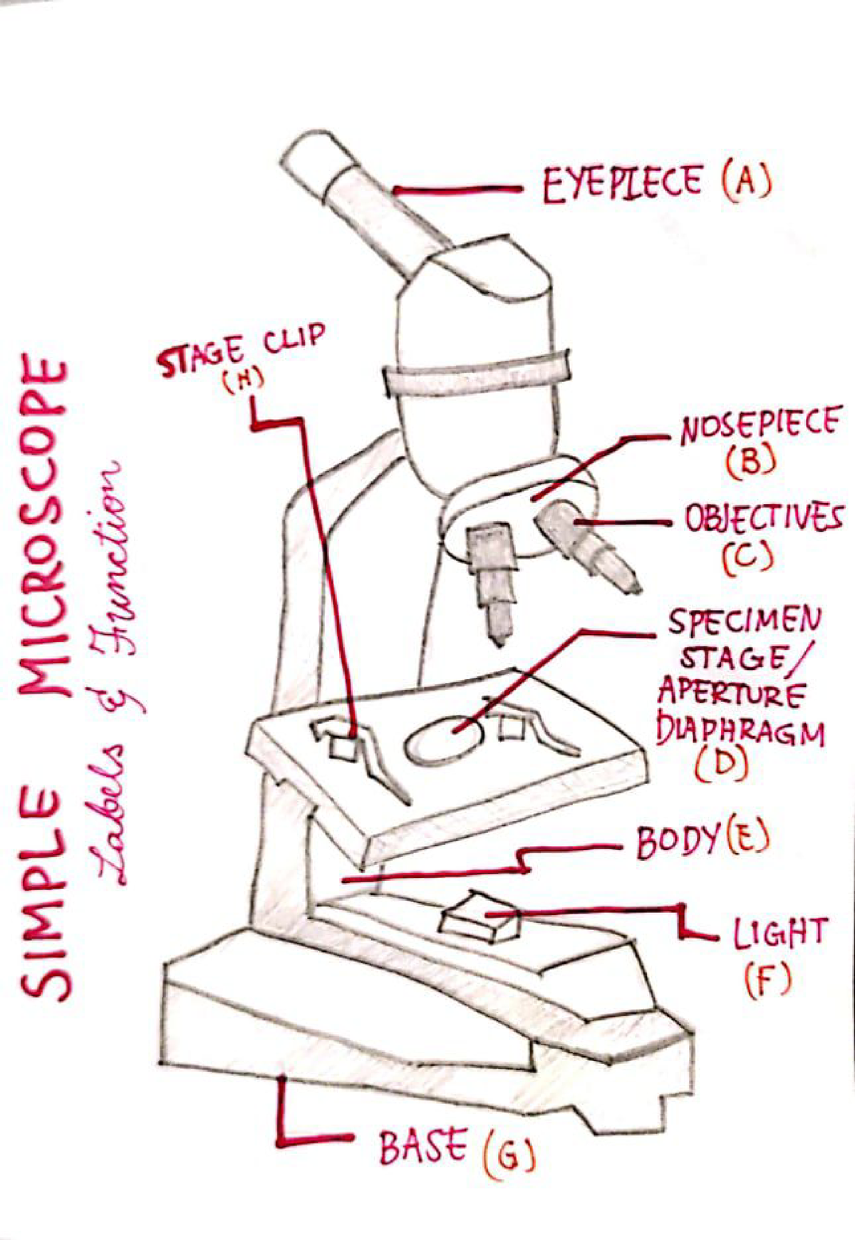
This is a common compound microscope. Label its parts from A ...
Microscope: Definition, Anatomy, Types and Uses - Embibe There microscope anatomy includes three structural parts, i.e. head, base, and arm. Head - This is also known as the body; it carries the optical parts in the upper part of the microscope.. Base - It acts as microscopes support.It also carries microscopic illuminators. Arms - The microscope arm connects the base and the head and the eyepiece tube to the microscope base.

Simple Microscope Diagram, Formula, Definition, Discoverd by
Animal Cell Diagram: Functions & Structure - Collegedunia Collegedunia Team. Animal cells are eukaryotic cells that are seen specifically in animal tissues. It is characterised by the absence of cell wall, with cell organelles enclosed within the membrane of the cell .They contain membrane-bound nuclei. The diagram of animal cell is beneficial in understanding the structure and functions of an animal.

Simple Microscope: Definition, working, diagram, properties, Uses
Fully Labelled Diagram Of A Toad - cms2.ncee.org Fully Labelled Diagram Of A A Study of the Microscope and its Functions With a Labeled Diagram. To better understand the structure and function of a microscope, we need to take a look at the labeled microscope diagrams of the compound and electron microscope. These diagrams clearly… The Structure of an Atom Explained With a Labeled Diagram ...
Junior cert Labelling Microscope - Labelled diagram
Microscope Types (with labeled diagrams) and Functions Has a higher level of magnification - Typically up to 2000x. Is used in hospitals and forensic labs by scientists, biologists and researchers to study micro organisms. Compound microscope labeled diagram. Compound microscope functions: It finds great application in areas of pathology, pedology, forensics etc.

Draw a neat labelled diagram of a compound microscope class ...
Plant Cell: Definition, Types of Plant Cells and More - Embibe Even though plant and animal cells are eukaryotic and share a few cell organelles, plant cells are quite distinct compared to animal cells as they perform different functions. These differences can be clearly understood when the cells are examined under an electron microscope. Observe the labelled diagram of plant cell structure as given below:

Living Environment Course
Bright-field microscope (Compound light microscope) - Diagram (Parts ... A few applications of the bright-field microscope include: Used to observe, analyze, and study plant cells. Used to view, magnify, and study about animal cells. Used to clearly study the morphologies of bacterial, and viral organisms. Also used in the study of parasites like paramecium. It finds use in agricultural laboratories to study soil ...

Label Microscope Diagram - EnchantedLearning.com
Compound Microscope - Diagram (Parts labelled), Principle and Uses See: Labeled Diagram showing differences between compound and simple microscope parts Structural Components. The three structural components include. 1. Head. This is the upper part of the microscope that houses the optical parts. 2. Arm . This part connects the head with the base and provides stability to the microscope.

A Study of the Microscope and its Functions With a Labeled ...
› techniques › multi-photonMultiphoton Microscopy | Nikon’s MicroscopyU This is a valuable enhancement to the capability of the conventional microscope since ultraviolet wavelengths below approximately 300 nanometers are very problematic for regular microscope optics. Higher-order non-linear effects, such as four-photon absorption, have been experimentally demonstrated, although it is unlikely that these phenomena ...

How to draw Microscope diagram for beginners - step by step

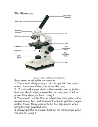
The Microscope

Microscope With Labels clip art | Microscope parts ...
Microscope Types (with labeled diagrams) and Functions
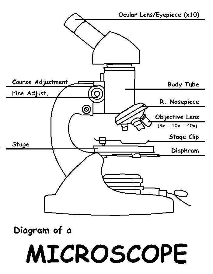
Diagram of a Microscope by ScienceDoodles on DeviantArt
MICROBIO 16 Parts of a Compound Microscope with Diagram and ...

Label the microscope — Science Learning Hub

Simple Microscope - Diagram (Parts labelled), Principle ...
Microscope labeled diagram
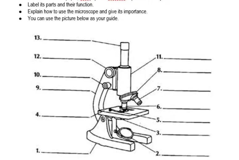
Answered: Label its parts and their function.… | bartleby

Can someone can send me diagram of this compound microscope ...

List: Parts of a Microscope and their Function | Pathwooded

Microscope Maintenance Tips | Science supplies, Multi step ...

Microscope diagram labeled | Clipart Panda - Free Clipart Images
Untitled Document

Microscope Labeling

Compound and Stereo- microscopes - Microscopes 4 Schools
Microscope- Simple-AND Compound-WITH- Label - BS in Education ...

Dissecting Stereo Microscope Parts and Functions
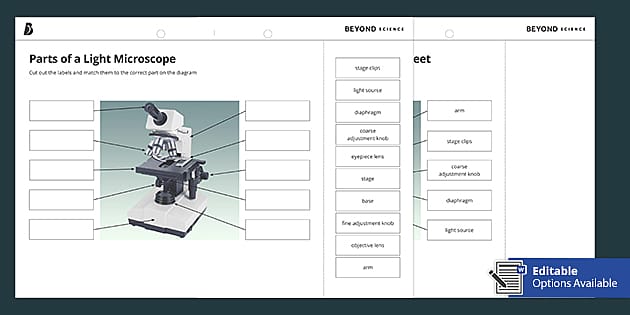
Parts of a Light Microscope Cut and Stick Worksheet - Twinkl

Microscopy: History, Types of Microscope, Diagram - Embibe

A labeled diagram of a microscope. MLT 101. :) | Teaching ...

Diagram of traveling microscope setup with implant cast and ...

Parts Of A Microscope Labeling Teaching Resources | TPT
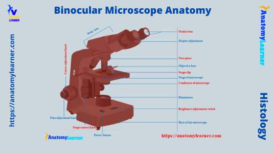
Binocular Microscope Anatomy - Parts and Functions with a ...
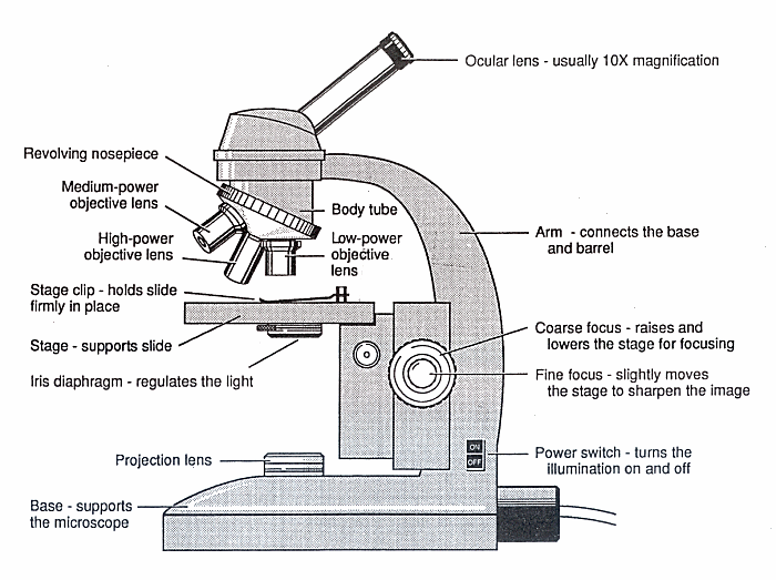
Diagram of a Microscope - Guide to using a microscope

Types of Microscopes: Definition, Working Principle, Diagram ...
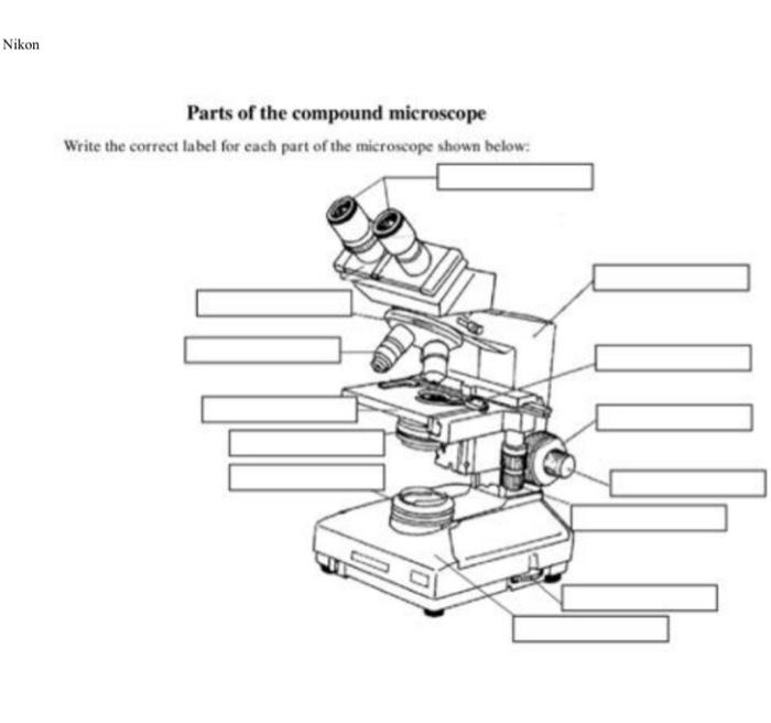
Solved Nikon Parts of the compound microscope Write the ...

Parts of a microscope with functions and labeled diagram
Compound Microscope Parts, Diagram Definition, Application ...

monocular labeling Diagram | Quizlet

Labeling the Parts of the Microscope | Microscope World Resources


Komentar
Posting Komentar