40 eye diagram not labeled
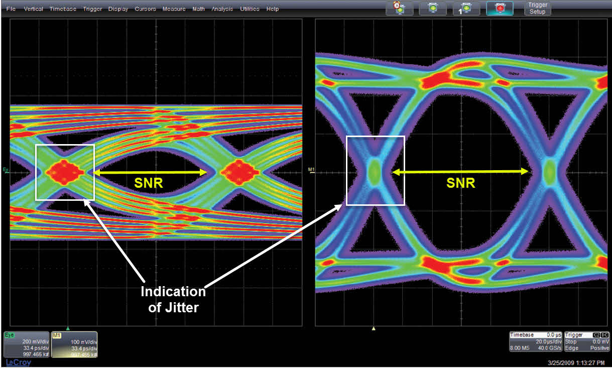
Labelling the eye — Science Learning Hub Use your mouse or finger to hover over a box to highlight the part to be named. Drag and drop the text labels onto the boxes next to the eye diagram If you want to redo an answer, click on the box and the answer will go back to the top so you can move it to another box. If you want to check your answers, use the 'Reset incorrect' button. What does an eye diagram or eye pattern on an oscilloscope mean? An eye diagram is a pattern shown on an oscilloscope that depicts a fuller view of what a digital signal stream looks like from a more holistic viewpoint, one could say. What's actually happening is that the oscilloscope is receiving, or sampling, a digital signal (a stream of step functions that represent 0s and 1s in varying patterns.)
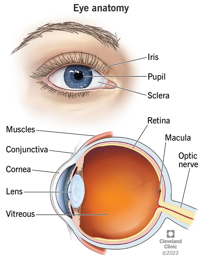
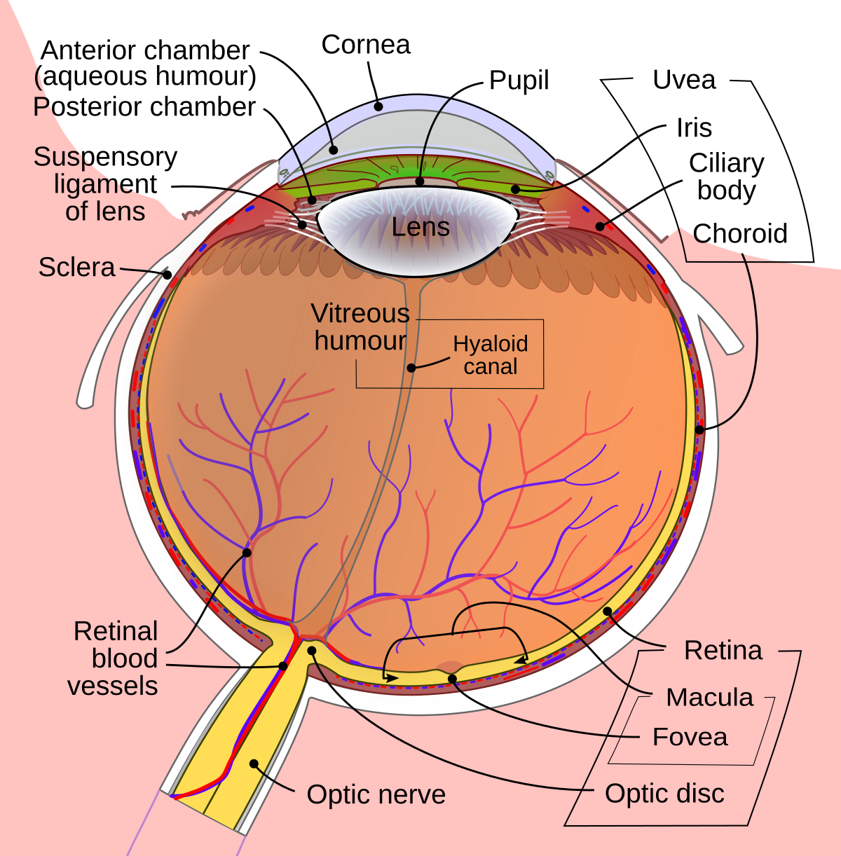
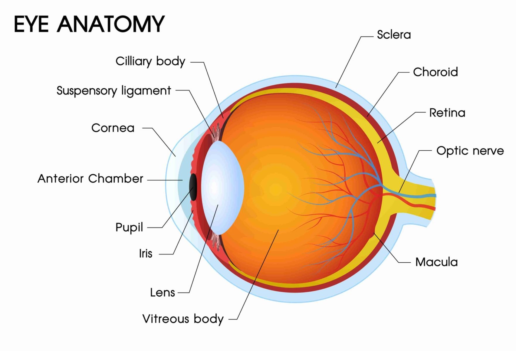
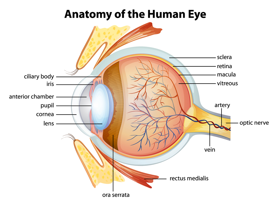
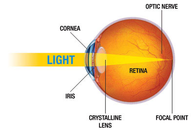
Eye Anatomy: Parts of the Eye and How We See Behind the anterior chamber is the eye's iris (the colored part of the eye) and the dark hole in the middle called the pupil. Muscles in the iris dilate (widen) or constrict (narrow) the pupil to control the amount of light reaching the back of the eye. Directly behind the pupil sits the lens. The lens focuses light toward the back of the eye.

Eye diagram not labeled
Labelling the eye — Science Learning Hub Labelling the eye. The human eye contains structures that allow it to perceive light, movement and colour differences. In this activity, students use online or paper resources to identity and label the main parts of the human eye. Citizen science. Teacher PLD. Unlabeled Skull Diagram The Splanchnocranium is the facial skeleton. The Neurocranium is the braincase. The skull in infants is made up of 45 separate elements but as an adult it is normally made up of 28 elements (including the ear ossicles) (White & Folkens 77). Jul 11, · Printable Eye Diagram Quiz Unlabeled on Diagram Site. This diagram pictures uploaded by ... Eye Anatomy: 16 Parts of the Eye & Their Functions - Vision Center The conjunctiva is the membrane covering the sclera (white portion of your eye). The conjunctiva also covers the interior of your eyelids. Conjunctivitis, often known as pink eye, occurs when this thin membrane becomes inflamed or swollen. Other eye disorders that affect the conjunctiva include:
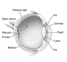
Eye diagram not labeled. Cow's Eye Dissection - Eye diagram - Exploratorium Learn how to dissect a cow's eye in your classroom. This resource includes: a step-by-step, hints and tips, a cow eye primer, and a glossary of terms. Cow's Eye Dissection - Eye diagram Lens of the Eye - All About Vision The lens of the eye, also called the crystalline lens, is an important part of the eye's anatomy that allows the eye to focus on objects at varying distances. It is located behind the iris and in front of the vitreous body. In its natural state, the lens looks like an elongated sphere — a shape known as ellipsoid — that resembles a ... Retina - Wikipedia The retina (from Latin: rete "net") is the innermost, light-sensitive layer of tissue of the eye of most vertebrates and some molluscs.The optics of the eye create a focused two-dimensional image of the visual world on the retina, which then processes that image within the retina and sends nerve impulses along the optic nerve to the visual cortex to create visual perception. PDF Parts of the Eye - National Institutes of Health Eye Diagram Handout Author: National Eye Health Education Program of the National Eye Institute, National Institutes of Health Subject: Handout illustrating parts of the eye Keywords: parts of the eye, eye diagram, vitreous gel, iris, cornea, pupil, lens, optic nerve, macula, retina Created Date: 12/16/2011 12:39:09 PM
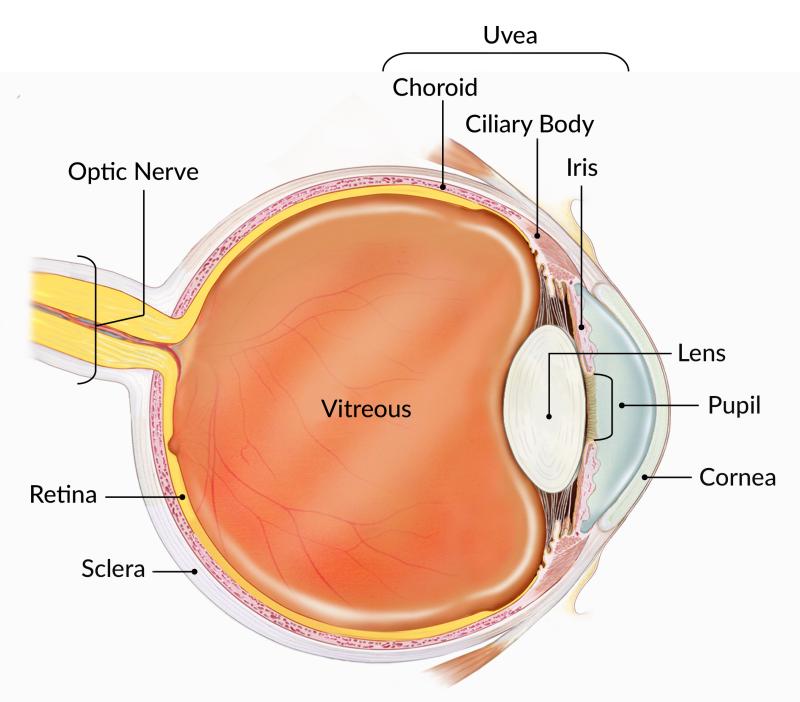
PDF Eye Anatomy Handout - National Institutes of Health Eye Anatomy Handout Author: National Eye Institute , National Eye Health Education Program Subject: Diabetes and Healthy Eyes Toolkit and Website Keywords: Eye anatomy, eye diagram, cornea, iris, lens, macula, optic nerve, pupil, retina, vitrous gel, diabetic eye disease. Created Date: 6/27/2012 11:57:40 AM Virtual 3D Eye Model | Johnson & Johnson Vision Virtual 3D Eye Model. Published on Oct 24, 2017. 5 Minutes Read. Learn basic eye anatomy with interactive models, view video tutorials of common refractive errors, and see how contact lenses can help give you clear vision. Viewing Normal. Eye Anatomy Diagram - EnchantedLearning.com Definitions : Aqueous humor - the clear, watery fluid inside the eye. It provides nutrients to the eye. Astigmatism - a condition in which the lens is warped, causing images not to focus properly on the retina. Binocular vision - the coordinated use of two eyes which gives the ability to see the world in three dimensions - 3D. Structure and Functions of Human Eye with labelled Diagram - BYJUS The External Structure of an Eye. Sclera: It is a white visible portion. It is made up of dense connective tissue and protects the inner parts. Conjunctiva: It lines the sclera and is made up of stratified squamous epithelium. It keeps our eyes moist and clear and provides lubrication by secreting mucus and tears.
Eye Diagram Printable: Free Worksheet for Kids Work with your child using this parts of the eye worksheet to help him or her learn more about their sense of sight! Completing this worksheet will help your child: • Identify different parts of the eye using an image • Match the words to the correct part of the eye • Gain new vocabulary Diagram of the Eye - Lions Eye Institute Instructions Click the parts of the eye to see a description for each. Hover the diagram to zoom. Need any help? If you would like to know more about us, or want to make an appointment, please don't hesitate to get in touch. (08) 9381 0777 carecentre@lei.org.au Request an appointment Customer Care Centre (08) 9381 0777 PDF Anatomy of an Eye Diagram - Tektronix Figure 6. An eye diagram triggered from a clock recovered from the data signal using a narrow loop bandwidth clock recovery scheme. Figure 5. An incomplete eye diagram formed by triggering on data. Figure 8. An eye diagram triggered such that the delay between jittered clock and jittered data destructively interferes. Figure 7. Atom: Definition, Structure & Parts with Labeled Diagram Oct 23, 2021 · An atom’s size is tiny, with a diameter of 0.1 to 0.5 nanometers (1 × 10 −10 to 5 × 10 −10 m). Thus they cannot be seen with our naked eye. A layer of an atom is somewhat similar to a sheet of paper.
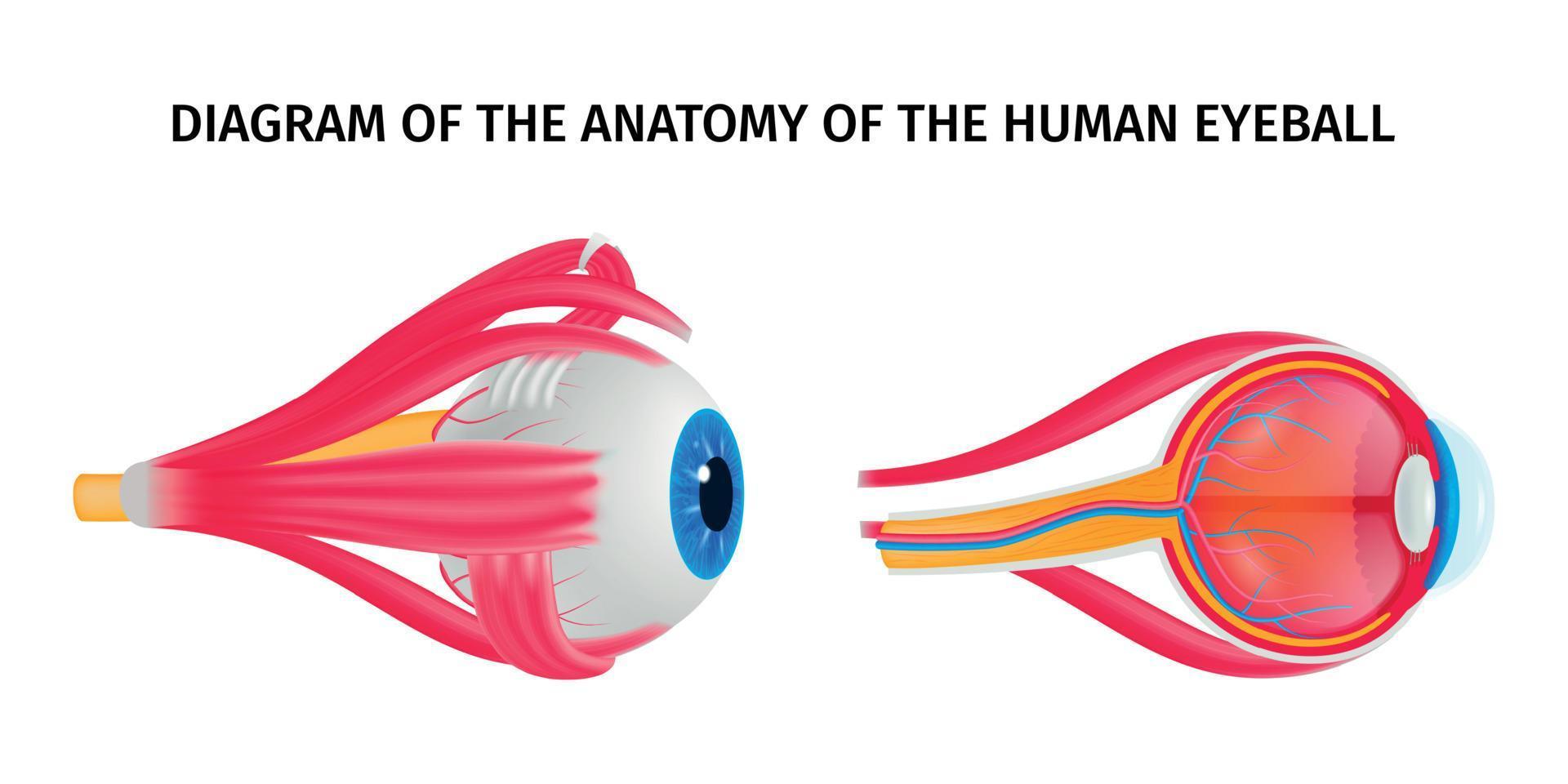
Human Eye Ball Anatomy & Physiology Diagram
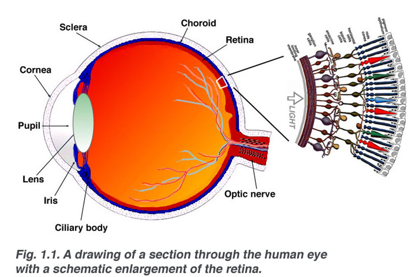
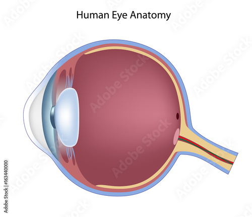
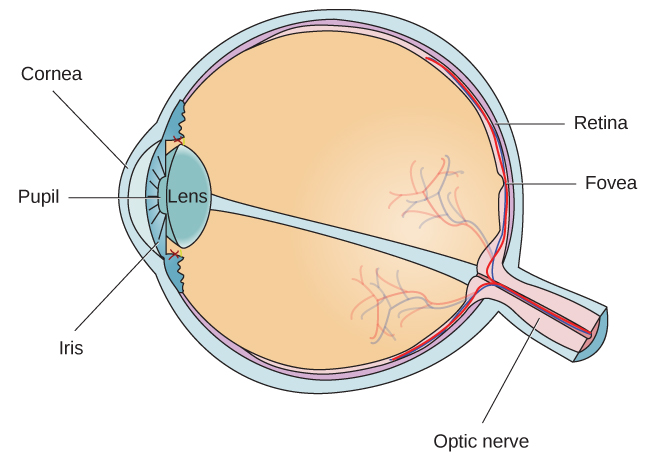
Vision and Eye Diagram: How We See - AARP Light reflects off the object we're looking at and enters the eye through the cornea, a clear, thin, dome-shaped tissue at the very front of the eye. The cornea has a curvature to it and covers the eye, kind of like a crystal covering the face of a watch. "When rays of light enter the eye, they're sort of parallel to each other," says Rosen.
Eyes: How They Work, Anatomy & Common Conditions
The Eye Diagram: What is it and why is it used? The eye diagram is used primarily to look at digital signals for the purpose of recognizing the effects of distortion and finding its source. To demonstrate using a Tektronix MDO3104 oscilloscope, we connect the AFG output on the back panel to an analog input channel on the front panel and press AFG so a sine wave displays. Then we press Acquire.

The Eye - Science Quiz
Eye Anatomy: A Closer Look At the Parts of the Eye - All About Vision Eye anatomy: A closer look at the parts of the eye. When surveyed about the five senses — sight, hearing, taste, smell and touch — people consistently report that their eyesight is the mode of perception they value (and fear losing) most. Despite this, many people don't have a good understanding of the anatomy of the eye, how vision works ...

Sensory Structures | BioNinja
The Eyes (Human Anatomy): Diagram, Optic Nerve, Iris, Cornea ... - WebMD Your eye is a slightly asymmetrical globe, about an inch in diameter. The front part (what you see in the mirror) includes: Iris: the colored part; Cornea: a clear dome over the iris; Pupil: the ...
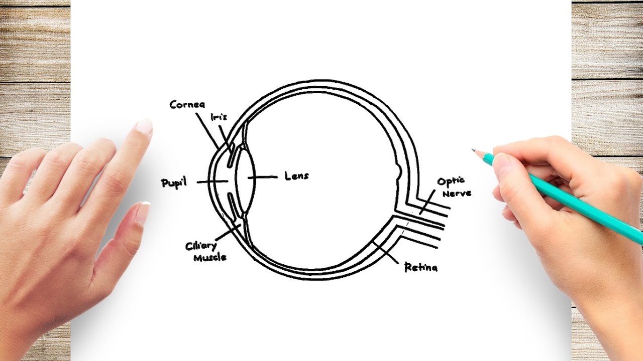
How to Draw Human Eye Diagram Easy Step
Course Help Online - Have your academic paper written by a ... Whether to reference us in your work or not is a personal decision. If it is an academic paper, you have to ensure it is permitted by your institution. We do not ask clients to reference us in the papers we write for them. When we write papers for you, we transfer all the ownership to you.
Structure and Function of the Eyes - Eye Disorders - MSD ...
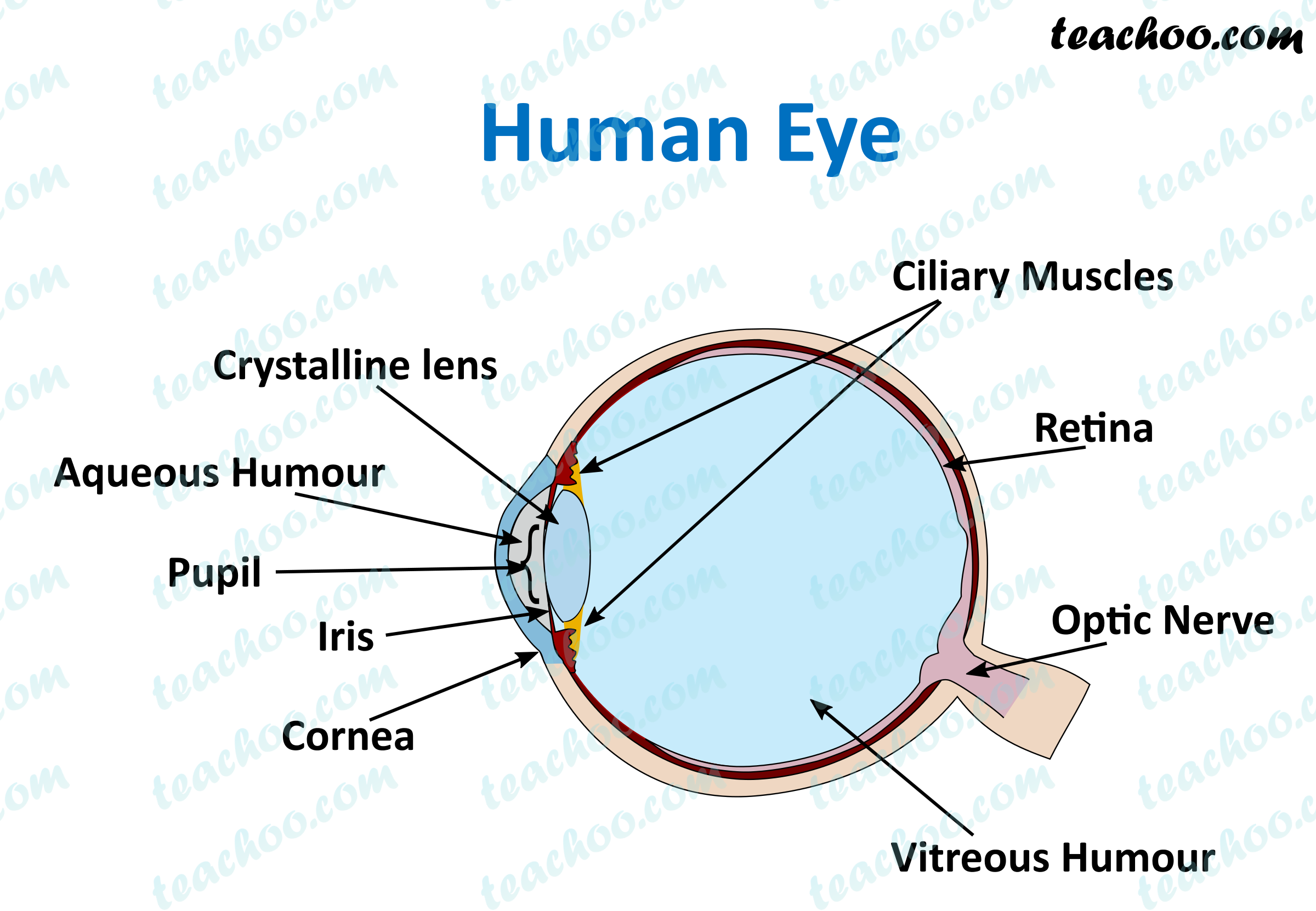
Labelled Diagram of Human Eye, Explanation and Function - VEDANTU The human eye is a part of the sensory nervous system. Labeled Diagram of Human Eye The eyes of all mammals consist of a non-image-forming photosensitive ganglion within the retina which receives light, adjusts the dimensions of the pupil, regulates the availability of melatonin hormones, and also entertains the body clock.

blank eye diagrams - Bing Images | Human eye diagram, Anatomy ...
Eye Test: 3 Free Eye Charts to Download and Print at Home Feb 27, 2019 · Not many humans have 20/10 vision or better, but some animals do. It's believed that most birds of prey have 20/5 acuity — or better. Eye chart limitations. Since eye charts only measure visual sharpness, they can help your eye doctor figure out whether you need prescription eyeglasses or contact lenses. They can also help your local ...
Simple Anatomy of the Retina by Helga Kolb – Webvision
Compound Microscope- Definition, Labeled Diagram, Principle ... Apr 03, 2022 · The naked eye can now view the specimen at magnification 400 times greater and so microscopic details are revealed. Alternatively, the magnification of the compound microscope is given by: m = D/ f o * L/f e

File:Eye Diagram without text.gif - Wikimedia Commons
Anatomy of an Eye Diagram: How to Construct & Trigger Mask testing is an abbreviated eye diagram test for the quick testing of transmitters in manufacturing. Rather than measuring all parametric aspects of the eye, mask testing defines key areas in the eye that are deemed to be "keep-out" areas - if any of the signal is detected to be in this region, then the device fails.
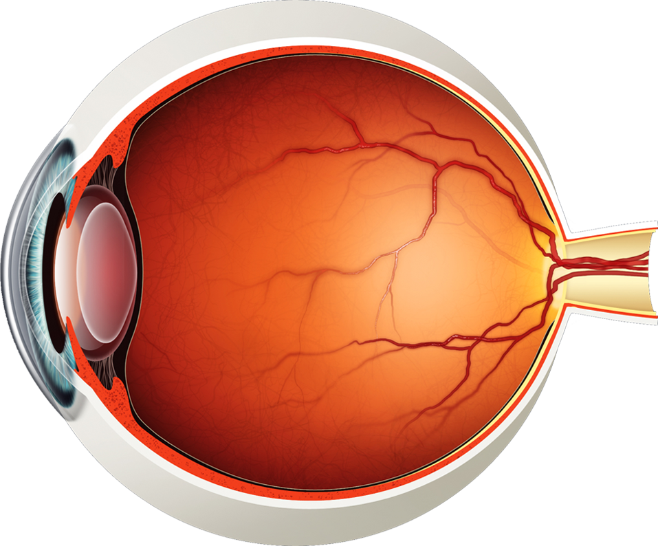
Diagram of the Eye - Lions Eye Institute
Eye Diagram Quiz - ProProfs Quiz Try this amazing Eye Diagram Quiz quiz which has been attempted 5391 times by avid quiz takers. Also explore over 72 similar quizzes in this category. Take Quizzes. Animal; Nutrition; ... Can you label the parts of the eye in the quiz below? Give it a try and evaluate yourself. The eye has many important parts, each with different functions ...
Eye in Cross Section : Anatomy : The Eyes Have It
Label Parts of the Human Eye - University of Dayton Parts of the Eye. Select the correct label for each part of the eye. The image is taken from above the left eye. Click on the Score button to see how you did. Incorrect answers will be marked in red. ...

Label Parts of the Human Eye
File:Diagram of human eye without labels.svg - Wikimedia This file is licensed under the Creative Commons Attribution-Share Alike 3.0 Unported license.: You are free: to share - to copy, distribute and transmit the work; to remix - to adapt the work; Under the following conditions: attribution - You must give appropriate credit, provide a link to the license, and indicate if changes were made. You may do so in any reasonable manner, but not in ...
How the Eyes Work | National Eye Institute
blank eye diagrams - Bing Images | Human eye diagram, Human ear diagram ... Inspiring Anatomy Human Ear Diagram Worksheet worksheet images. Holly Crimm. School. Human Skeleton Anatomy. Gross Anatomy. Heart Anatomy. Radiology Student. ... This is a quiz called Label the Eye and was created by member LegoA1. Emily Iboa. Apologia General Science. Human Heart Diagram. Medical Drawings.

How the eye works | RNIB
Eye Diagram - an overview | ScienceDirect Topics An eye diagram provides a simple and useful tool to visualize intersymbol interference between data bits. Figure 24a shows a perfect eye diagram. A square bit stream (i.e., series of symbol '1's and '0's) is sliced into sub-bit stream with predetermined eye intervals (i.e., several bit periods), and displayed through bit analyzing equipment (e.g., digital channel analyzer), overlapping ...
Eye Health: Anatomy of the Eye - VisionAware
Allergy - Wikipedia A major breakthrough in understanding the mechanisms of allergy was the discovery of the antibody class labeled immunoglobulin E (IgE). IgE was simultaneously discovered in 1966–67 by two independent groups: [135] Ishizaka 's team at the Children's Asthma Research Institute and Hospital in Denver, USA, [136] and by Gunnar Johansson and Hans ...
Eye - Wikipedia
Eye diagram basics: Reading and applying eye diagrams - EDN Figure 3 (a) Improper termination causes an eye diagram to look stressed. (b) Proper termination relaxes the eye. As can be seen in Figure 4, an eye diagram can reveal important information. It can indicate the best point for sampling, divulge the SNR (signal-to-noise ratio) at the sampling point, and indicate the amount of jitter and distortion.
Human Eye Anatomy Diagram 5751327 Vector Art at Vecteezy
Eye Diagram (EYE) - INTERCONNECT Element - Ansys Optics The Eye Diagram element can also be used as a decoder when the reference signal/bit pattern is provided. Note that in this case, the Eye Diagram element will recover the signal based on the input bit pattern, so it is possible to recover level one signal in a lower power level than the level zero signal when the modulator swaps the signal ...
Anatomy of the eye, non-labeled Stock Illustration | Adobe Stock
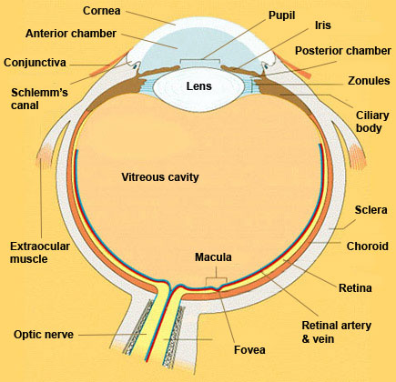
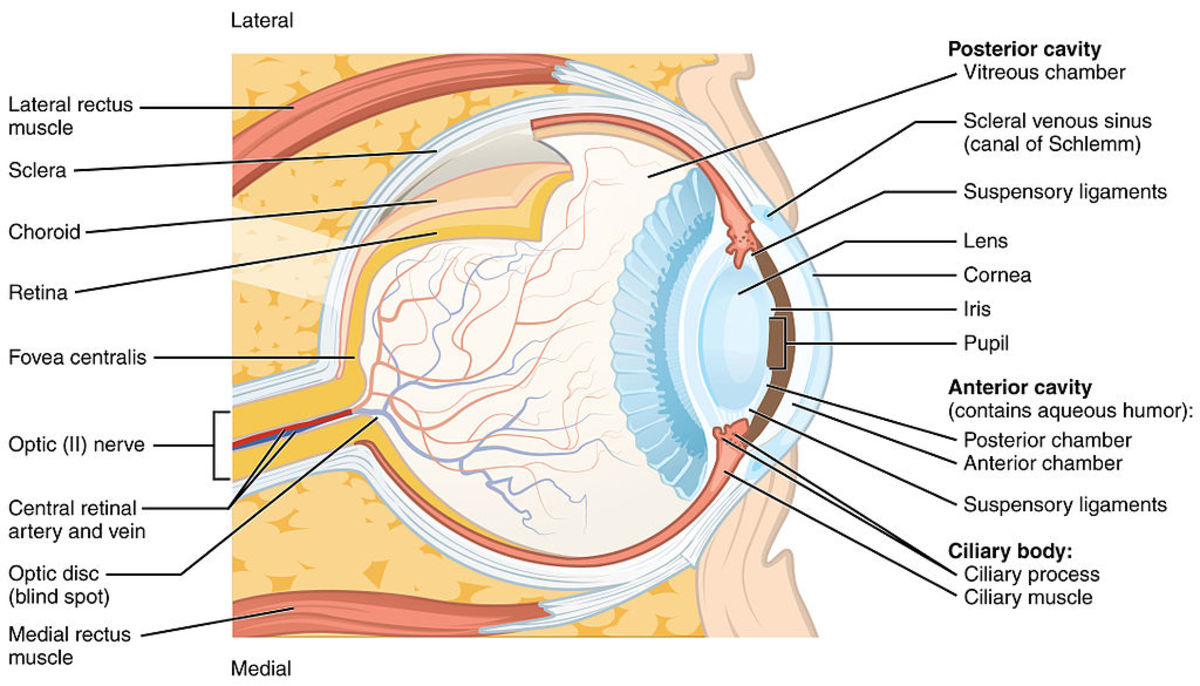
Eye Diagram With Labels and detailed description - BYJUS A brief description of the eye along with a well-labelled diagram is given below for reference. Well-Labelled Diagram of Eye The anterior chamber of the eye is the space between the cornea and the iris and is filled with a lubricating fluid, aqueous humour. The vascular layer of the eye, known as the choroid contains the connective tissue.

Schematic drawing of the human eye. Adapted from ...
Eye Anatomy: 16 Parts of the Eye & Their Functions - Vision Center The conjunctiva is the membrane covering the sclera (white portion of your eye). The conjunctiva also covers the interior of your eyelids. Conjunctivitis, often known as pink eye, occurs when this thin membrane becomes inflamed or swollen. Other eye disorders that affect the conjunctiva include:
Label Eye Printout - EnchantedLearning.com
Unlabeled Skull Diagram The Splanchnocranium is the facial skeleton. The Neurocranium is the braincase. The skull in infants is made up of 45 separate elements but as an adult it is normally made up of 28 elements (including the ear ossicles) (White & Folkens 77). Jul 11, · Printable Eye Diagram Quiz Unlabeled on Diagram Site. This diagram pictures uploaded by ...
Eye Anatomy Diagram - EnchantedLearning.com
Labelling the eye — Science Learning Hub Labelling the eye. The human eye contains structures that allow it to perceive light, movement and colour differences. In this activity, students use online or paper resources to identity and label the main parts of the human eye. Citizen science. Teacher PLD.
Eye Anatomy: The 9 Main Parts of the Eye | Specialty Eye ...

Eye Anatomy (labelled), illustration - Stock Image - C043 ...

human eye | Definition, Anatomy, Diagram, Function, & Facts ...
What does an eye diagram or eye pattern on an oscilloscope mean?
![Cross sectional diagram of human eye [1]. | Download ...](https://www.researchgate.net/publication/276541864/figure/fig1/AS:612895498964992@1523137082339/Cross-sectional-diagram-of-human-eye-1.png)
Cross sectional diagram of human eye [1]. | Download ...
Anatomy and Structure of the Human Eye (With Diagrams ...
Eye Anatomy - Exeter Eye
6,889 Human Eye Diagram Images, Stock Photos & Vectors ...
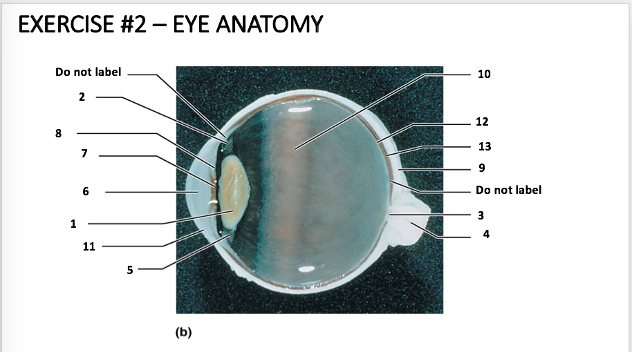
Solved EXERCISE #2 – EYE ANATOMY Do not label 10 2 12 8 13 7 ...
How We See | Introduction to Psychology

Jarvis: Chapter 14: Eyes Diagram | Quizlet
How the Human Eye Works | Cornea Layers/Role | Light Rays
/GettyImages-695204442-b9320f82932c49bcac765167b95f4af6.jpg)
Structure and Function of the Human Eye

How the eyes work - Perkins School for the Blind

Q10 Draw a labeled sketch of the human eye...

The Eye - diagram to label | Teaching Resources
Human Eye - Different Parts and their functions - Class 10 ...

Eye Anatomy - Bell Booth Sirkka Fabris Optometrists

eye label Diagram | Quizlet


Komentar
Posting Komentar