40 clam diagram labeled
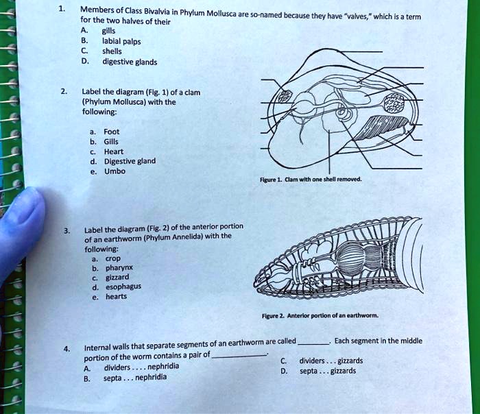
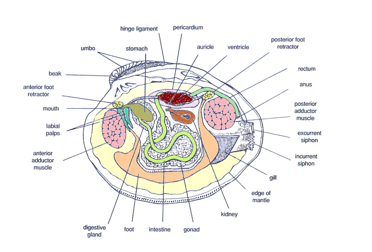
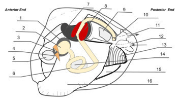
Solved C. Identify each labeled structure on the clam - Chegg C. Identify each labeled structure on the clam diagram. Write your answers in the chart below the diagram and briefly state the function of each structure. Here is a list of structures you are looking for: shell, adductor muscles, incurrent siphon, excurrent siphons, foot, gills, mantle, mouth, labial palps, heart, and anus. opengeology.org › textbook › 7-geologic-time7 Geologic Time – An Introduction to Geology In the block diagram, the sequence of geological events can be determined by using the relative-dating principles and known properties of igneous, sedimentary, metamorphic rock (see Chapter 4, Chapter 5, and Chapter 6). The sequence begins with the folded metamorphic gneiss on the bottom. Next, the gneiss is cut and displaced by the fault ...
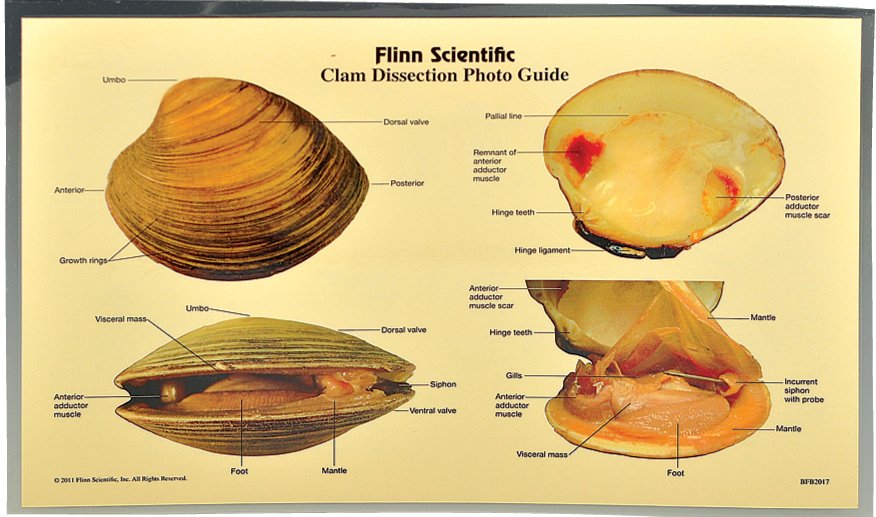
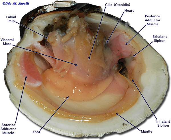
DOC Clam Dissection - wallingford.k12.ct.us Continue following the intestine toward the posterior end of the clam. Find the . anus. just behind the posterior adductor muscle. Use your probe to trace the path of food & wastes from the incurrent siphon through the clam to the excurrent siphon. Answer the questions on your lab report & label the diagrams of the internal structures of the clam.

Clam diagram labeled
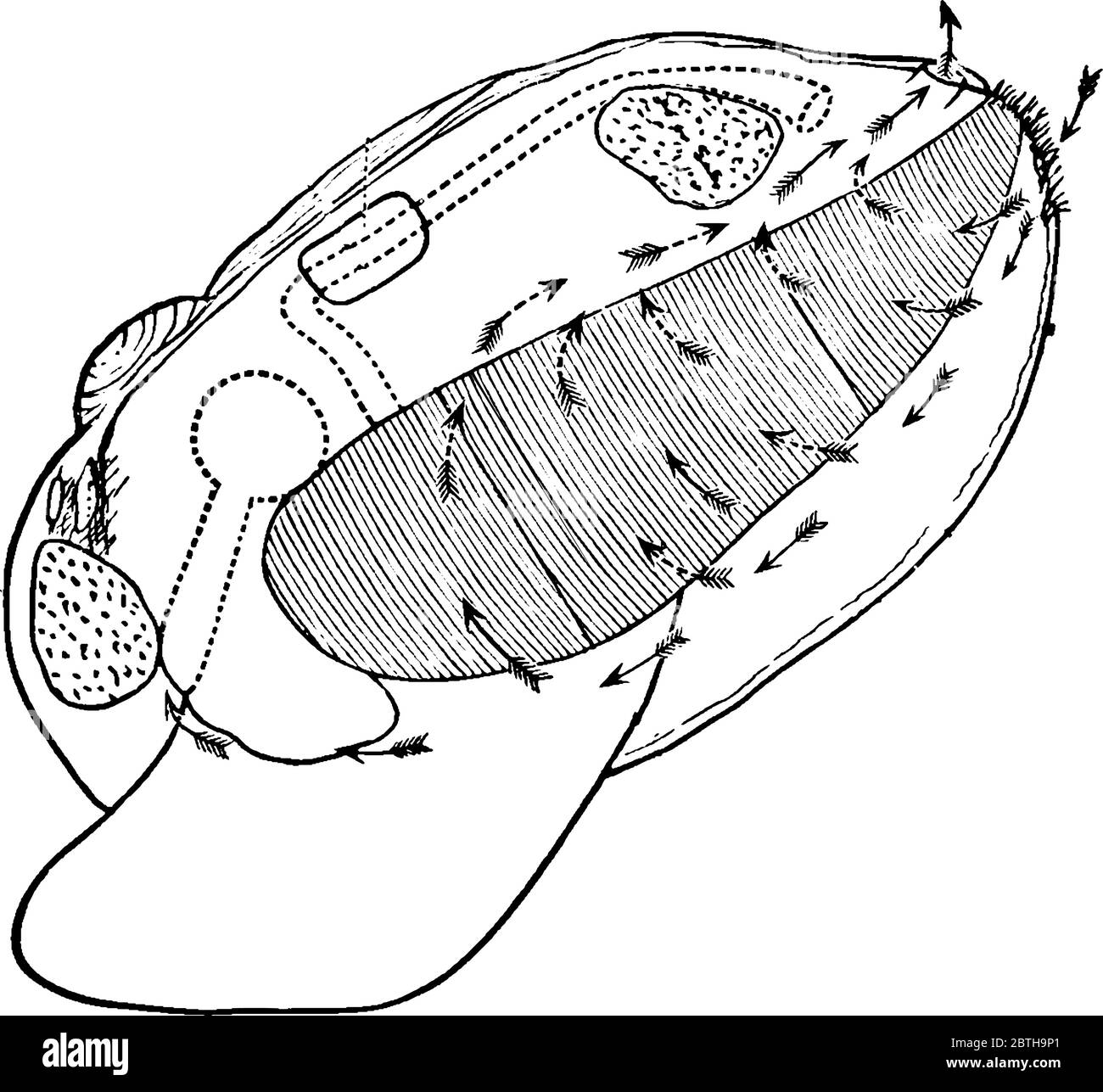
PDF Anatomy of a Clam Ask the students to label and/or draw (if using lab notebooks) each step of the dissection and identify major organs and their uses (information will be in dissection guide). Post work/Clean-up: 1. When students are finished with the dissection, have them fold all materials into their paper towels and set aside a separate trashcan for dissection PDF Clam (Mercenaria mercenaria) Dissection - Muse TECHNOLOGIES anatomy of a clam. 3.Locate the umbo, the bump at the anterior end of the valve. This is the oldest part of the clam shell. ... Answer the questions on your lab report and draw and label (in your sketchbook) a diagram of the internal anatomy of the clam. Use the arrows on the clam diagram to trace the pathway of food as it clam anatomy Diagram | Quizlet Start studying clam anatomy. Learn vocabulary, terms, and more with flashcards, games, and other study tools.
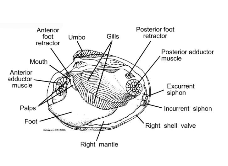
Clam diagram labeled. wls.dubaishopcheznous.fr › shakespeare-gx235Shakespeare gx235 manual shakespeare gx235 manual. mphil dphil in economics. health lottery login.The long read: DNP is an industrial chemical used in making explosives. If swallowed, it can cause a horrible death - and yet it is still being aggressively marketed to vulnerable people online. Clam Diagram Labeled Diagram - See attached diagram. Label the Parts of the Clam Marine Biology, Clams, Science Activities, Juices,. Visit. Clam Anatomy Labeling Page .. Clam Anatomy Coloring Page. Explain the functions of the organs of the clam (Anodonta). Diagrams and Key: From Biodidac: Clam in Color. Structures to pin and label: 1. excurrent siphon, 2. › searchImages, Stock Photos & Vectors | Shutterstock Jun 30, 2022 · Find stock images in HD and millions of other royalty-free stock photos, illustrations and vectors in the Shutterstock collection. Thousands of new, high-quality pictures added every day. nuvqb.grajnisko.pl › double-bluff-county-parkSearch Icon - nuvqb.grajnisko.pl BIDS: Bids must be legibly printed or type written, labeled "Double Chip Seal Road Surfacing Project - Ellison Bluff County Park", and submitted electronically (to [email protected] or sealed in an opaque envelope plainly marked "Double Chip Seal Road Surfacing Project - Ellison Bluff County Park" and mailed or delivered to the Facilities ...
Clam Dissection - BIOLOGY JUNCTION Leave the tip of the screwdriver between the valves and place the clam in the pan with the left valve up. Locate the adductor muscles. With your blade pointing toward the dorsal edge, slide your scalpel between the upper valve & the top tissue layer. Cut down through the anterior adductor muscle, cutting as close to the shell as possible. up.codes › viewer › new_york_cityChapter 5: Exhaust Systems, NYC Mechanical Code 2014 | UpCodes The switch shall be a break-glass or other approved type and shall be labeled: VENTILATION SYSTEM EMERGENCY SHUTOFF. The exhaust ventilation shall be designed to consider the density of the potential fumes or vapors released. For fumes or vapors that are heavier than air, exhaust shall be taken from a point within 12 inches (305 mm) of the floor. mussel internal anatomy clam labeled dissection dissected intestine anus lab mussel anatomy mouth muscle posterior bivalves mantle adductor unlabeled butterflied foot dorsal anterior Mussels mussel mussels mytilus diagram parts edulis body modiolus horse edible common showing invertebrates bumblebee Phylum - Mollusca (Gastropods, Bivalves, Cephalopods) New Page 1 [ez002.k12.sd.us] Leave the tip of the screwdriver between the valves and place the clam in the pan with the left valve up. 6. Locate the adductor muscles. With your blade pointing toward the dorsal edge, slide your scalpel or scissors between the upper valve & the top tissue layer.
Clam Dissection Diagram | Quizlet Start studying Clam Dissection. Learn vocabulary, terms, and more with flashcards, games, and other study tools. Scheduled maintenance: Saturday, September 10 from 11PM to 12AM PDT. ... ANATOMY AND PHYSIOLOGY. Why does the blind spot from the optic disc in either eye not result in a blind spot in the visual field? Verified answer. PDF Clam Dissection Guideline - Monadnock Regional High School 12. Observe the muscular foot of the clam ventral to the gills. It is attached to the soft visceral mass, which contains the other organs. Note the hatchet shape of the foot used to burrow into mud or sand. 13. Locate the palps, (mouth flaps) structures that surround & guide food into the clam's mouth.Beneath the palps, Clam Anatomy Labeling Page - Exploring Nature Clam Anatomy Labeling Page. Higher Resolution PDF for Printing. Click Here. Use Teacher Login to show answer keys or other teacher-only items. Link to More Info About this Animal (with Labeled Body Diagram) Click Here. Citing Research References. When you research information you must cite the reference. Citing for websites is different from ... Clam Anatomy Diagram Anatomy note Odysee Channel, Please Subscribe to Support. We are pleased to provide you with the picture named Clam Anatomy Diagram. We hope this picture Clam Anatomy Diagram can help you study and research. for more anatomy content please follow us and visit our website: . Anatomynote.com found Clam Anatomy Diagram from ...
en.wikipedia.org › wiki › Logan_International_AirportLogan International Airport - Wikipedia Runway 14/32, Logan's first major runway addition in more than forty years, opened on November 23, 2006. It was proposed in 1973, but was delayed in the courts. According to Massport records, the first aircraft to use the new airstrip was a Continental Express ERJ-145 regional jet landing on Runway 32, on the morning of December 2, 2006.
Clam Anatomy Diagram | Quizlet large muscles that hold the shell together. heart. A hollow, muscular organ that pumps blood throughout the body. muscular foot. used for movement. mantle. secretes mother of pearl; surrounds and protects the soft body of molluscs. palp. moves food particles to mouth in molluscs.
Bivalve (Clam) Diagram Quiz - PurposeGames.com About this Quiz. This is an online quiz called Bivalve (Clam) Diagram. There is a printable worksheet available for download here so you can take the quiz with pen and paper. This quiz has tags. Click on the tags below to find other quizzes on the same subject. biology.
Clam Diagram Quiz - By dwhite298 - Sporcle Top Contributed Quizzes in Science. 1. Almost a Science Sorting Gallery. 2. The First Ten... Nations in Space. 3. Medical Terminology Roots. 4.
Solved 4 5 7 6 This is a diagram of a dissected clam. | Chegg.com Biology questions and answers. 4 5 7 6 This is a diagram of a dissected clam. [Blank #1] To what phylum do these organisms belong? (Blank #2] Name the structure labeled 4. [Blank #3] Name the structure labeled 5. (hint: this one of 2 structures that hold the shells closed when you try to pry them open) [Blank #4] Name the structure labeled 6.
PDF Investigation #5 - Clam Anatomy - COSEE Locate the following parts of your clam according to the diagram: adductor muscles gills mantle excurrent siphon incurrent siphon stomach mouth foot intestine Lift the gills to find the stomach and intestines. Insert the skewer into the mouth and see that it empties into the stomach. Locate the foot that is used for digging.
PDF Taxonomy, Anatomy, and Biology of the Hard Clam Internal Clam 1 Mantle Shell Anatomy • Covers visceral or body mass • Holds in fluid • Secrets new shell 2. Ant. adductor muscle 3. Post adductor musclePost. adductor muscle • Hold valves shut 4. Pericardium cavity • Region covered with thin Region covered with thin, dark membrane • Contains 2-chambered heart and kidney in a fluid-filled sac 5.
opengeology.org › textbook › 5-weathering-erosion5 Weathering, Erosion, and Sedimentary Rocks – An ... A simplified soil profile, showing labeled layers. O Horizon: The top horizon is a thin layer of predominantly organic material, such as leaves, twigs, and other plant parts that are actively decaying into humus. A Horizon: The next layer, called topsoil, consists of humus mixed with mineral sediment. As precipitation soaks down through this ...
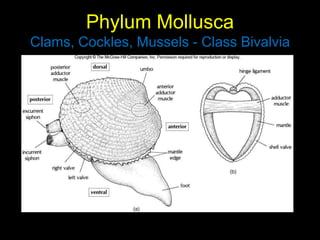
Clam Diagram & Parts | What Is a Clam? | Study.com Belonging to a diverse group of animals known as bivalves, clams can be identified by the presence of two valves, or shells, joined by a hinge that allows the two shells to open or close. A...
clam anatomy Diagram | Quizlet Start studying clam anatomy. Learn vocabulary, terms, and more with flashcards, games, and other study tools.
PDF Clam (Mercenaria mercenaria) Dissection - Muse TECHNOLOGIES anatomy of a clam. 3.Locate the umbo, the bump at the anterior end of the valve. This is the oldest part of the clam shell. ... Answer the questions on your lab report and draw and label (in your sketchbook) a diagram of the internal anatomy of the clam. Use the arrows on the clam diagram to trace the pathway of food as it
PDF Anatomy of a Clam Ask the students to label and/or draw (if using lab notebooks) each step of the dissection and identify major organs and their uses (information will be in dissection guide). Post work/Clean-up: 1. When students are finished with the dissection, have them fold all materials into their paper towels and set aside a separate trashcan for dissection












Komentar
Posting Komentar